This article is more than 1 year old
Take a look at atom’s shadow
Griffith University boffins ‘see’ ytterbium
This image is special, according to Griffith University: it’s the first time anyone’s captured the shadow of a single ytterbium atom, at optical wavelengths (images of single atoms are much easier to capture using scanning electron microscopes).
The image takes the world to the “extreme limit” of optical microscopy, the researchers say.
One of Australia’s hot-spots of quantum research, the Griffith team has been working on a technique to apply absorption imaging at the atomic scale for some years. A big part of its research has been in developing phase Fresnel lenses to collect the light in the image.
Absorption imaging is a technique that works at a host of scales, all the way up to the galactic, allowing astronomers (for example) to work out the characteristics of gas clouds by observing the light they absorb.
The Griffith researchers captured the shadow of an ytterbium ion by ion-trapping it (that is, using electrical fields to hold it in place), cooling it to a few millikelvin, and illuminating it at 369.5 nm wavelength. The light that passed through the ion was collected and focused through the phase Fresnel lens to produce the absorption image.
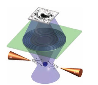
The experimental setup: tungsten electrodes trap the atom, which is illuminated
at 369.5 nm. The phase Fresnel lens focuses the image to a 4.8-micron spot.
Source: Nature Communications
The lens focused the image to a spot on a detector just 4.8 microns in size. This fine focus, according to Professor Dave Kielpinski of Griffith’s Centre for Quantum Dynamics, made the image darker and the shadow more visible.
The light illuminating the atom has to be controlled with superhuman precision. According to Daily Disruption, if the wavelength drifts by one part per billion, the image is lost.
“By using the ultra hi-res microscope we were able to concentrate the image down to a smaller area than has been achieved before, creating a darker image which is easier to see”, Professor Kielpinski said.
The high contrast achieved by the researchers is important for biomicroscopy, since it reduces the amount of light needed to illuminate very small samples – and therefore reduces the risk that the light may damage the sample. The same holds true for X-ray microscopy, the researchers say.
The research has been published in Nature Communications (abstract here).®
